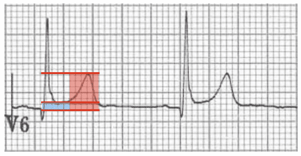
Saddle Shaped St Elevation - Dr. Smith's ECG Blog: A Very Subtle LAD Occlusion....T ... - The st segment starts from the j point (termination of qrs complex and the beginning of st segment) and ends with the t wave.
Saddle Shaped St Elevation - Dr. Smith's ECG Blog: A Very Subtle LAD Occlusion....T ... - The st segment starts from the j point (termination of qrs complex and the beginning of st segment) and ends with the t wave.. Its presence must be explained (there is no nonspecific st elevation). In pericarditis, this saddle shape is characteristically seen globally throughout the ecg. Saddle shaped st elevation across multiple leads. 1600 x 1188 png 2050kb. Prompt diagnosis of acute st segment elevation myocardial infarction (stemi) by the initial ecg is important in order to perform an urgent coronary angiography as soon as possible and achieve successful revascularization, therewith improving mortality and morbidity.
This page is about st segment j point elevation,contains ecg interpretation: Saddle shaped st elevation/ concave st elevation. • left ventricular aneurysm o persistent st elevation in leads that look at the affected area following an mi injury is shown on the ecg as an elevated st segment. Pr segment elevation or depression in patients with myocardial infarction indicates concomitant atrial ischaemia or infarction. Evaluation of st segment elevation criteria for the prehospital electrocardiographic diagnosis fo acute myocardial infarction.
Ecg 101 with answers from image.slidesharecdn.com The presence of a type 1 pattern with at least one clinial criterion is diagnostic of brugada syndrome. Usually after a viral illness. Serum sodium returned to normal after 2 weeks. Ecg patterns • rbbb • st elevations limited to right precordial leads v1 and v2 • saddle shaped or coved shaped st. Tachypnoea, tachycardia, hypoxia, haemoptysis, non specific st and t wave changes in anterior chest leads most common ecg finding, 'classical' s1q3t3 pattern is uncommon. Prompt diagnosis of acute st segment elevation myocardial infarction (stemi) by the initial ecg is important in order to perform an urgent coronary angiography as soon as possible and achieve successful revascularization, therewith improving mortality and morbidity. There is also st elevation in avl with st straightening in lead i. This page is about st segment j point elevation,contains ecg interpretation:
St elevation refers to a finding on an electrocardiogram wherein the trace in the st segment is abnormally high above the baseline.
• left ventricular aneurysm o persistent st elevation in leads that look at the affected area following an mi injury is shown on the ecg as an elevated st segment. Its presence must be explained (there is no nonspecific st elevation). The most likely ecg finding in this patient is sinus tachycardia. Lead ii is equally biphasic interpretation: Pericarditis is also associated with a concave st morphology; To explore more similar hd image on pngitem. St elevation is concave and no more than 5mm from j point. The st segment represents completed ventricular myocardial depolarization. Variable shapes of st segment elevations in ami goldberger al. Tachypnoea, tachycardia, hypoxia, haemoptysis, non specific st and t wave changes in anterior chest leads most common ecg finding, 'classical' s1q3t3 pattern is uncommon. Saddle shaped st elevation/ concave st elevation. Most cases are mild, and it typically resolves within a few weeks. There is also st elevation in avl with st straightening in lead i.
Sign up for the art's cyclery mailing list to get the latest sale information, special deals, and how to videos and articles that our customers love! Qrs complexes and t waves remain unchanged. Following pericardiocentesis there was a marked diuresis; There is st depression in the inferior leads, ii, iii, and avf. Its presence must be explained (there is no nonspecific st elevation).
Mimics of ST Elevation Myocardial Infarction (STEMI ... from www.ahcmedia.com T wave changes and q waves rarely seen in acute pericarditis. • left ventricular aneurysm o persistent st elevation in leads that look at the affected area following an mi injury is shown on the ecg as an elevated st segment. St elevation is concave and no more than 5mm from j point. There is st depression in the inferior leads, ii, iii, and avf. Pericarditis is also associated with a concave st morphology; St elevation refers to a finding on an electrocardiogram wherein the trace in the st segment is abnormally high above the baseline. Following pericardiocentesis there was a marked diuresis; Most cases show st elevation in both limb and chest leads.
Echo demonstrated a large pericardial effusion causing marked tamponade.
Saddle shaped st elevation/ concave st elevation. In pericarditis, this saddle shape is characteristically seen globally throughout the ecg. The st segment represents completed ventricular myocardial depolarization. Its presence must be explained (there is no nonspecific st elevation). T wave changes and q waves rarely seen in acute pericarditis. Diagnosing acute myoardial infarction (stemi) and 17 important differential diagnoses. This page is about st segment j point elevation,contains ecg interpretation: Qrs complexes and t waves remain unchanged. Learn all about st elevations (elevated st segments) on ecg; Most cases are mild, and it typically resolves within a few weeks. Sign up for the art's cyclery mailing list to get the latest sale information, special deals, and how to videos and articles that our customers love! Saddle shaped st elevation across multiple leads. Ecg patterns • rbbb • st elevations limited to right precordial leads v1 and v2 • saddle shaped or coved shaped st.
In type 2 and type 3 brugada syndrome, the st segment elevation is saddleback shaped (panels b and c, figure 10). The most likely ecg finding in this patient is sinus tachycardia. Prompt diagnosis of acute st segment elevation myocardial infarction (stemi) by the initial ecg is important in order to perform an urgent coronary angiography as soon as possible and achieve successful revascularization, therewith improving mortality and morbidity. Variable shapes of st segment elevations in ami goldberger al. Pericarditis is also associated with a concave st morphology;
Pericarditis ECG Changes • LITFL • ECG Library Diagnosis from litfl.com Tachypnoea, tachycardia, hypoxia, haemoptysis, non specific st and t wave changes in anterior chest leads most common ecg finding, 'classical' s1q3t3 pattern is uncommon. Ecg patterns • rbbb • st elevations limited to right precordial leads v1 and v2 • saddle shaped or coved shaped st. Lead ii is equally biphasic interpretation: Evaluation of st segment elevation criteria for the prehospital electrocardiographic diagnosis fo acute myocardial infarction. Following pericardiocentesis there was a marked diuresis; St elevation results from a prolonged lack of blood supply. Echo demonstrated a large pericardial effusion causing marked tamponade. Variable shapes of st segment elevations in ami goldberger al.
Echo demonstrated a large pericardial effusion causing marked tamponade.
In type 2 and type 3 brugada syndrome, the st segment elevation is saddleback shaped (panels b and c, figure 10). Most cases are mild, and it typically resolves within a few weeks. Diagnosing acute myoardial infarction (stemi) and 17 important differential diagnoses. T wave changes and q waves rarely seen in acute pericarditis. St elevation is concave and no more than 5mm from j point. Variable shapes of st segment elevations in ami goldberger al. Evaluation of st segment elevation criteria for the prehospital electrocardiographic diagnosis fo acute myocardial infarction. There is also st elevation in avl with st straightening in lead i. Learn all about st elevations (elevated st segments) on ecg; Prompt diagnosis of acute st segment elevation myocardial infarction (stemi) by the initial ecg is important in order to perform an urgent coronary angiography as soon as possible and achieve successful revascularization, therewith improving mortality and morbidity. In pericarditis, this saddle shape is characteristically seen globally throughout the ecg. Pericarditis is also associated with a concave st morphology; Saddle shaped st elevation/ concave st elevation.
You have just read the article entitled Saddle Shaped St Elevation - Dr. Smith's ECG Blog: A Very Subtle LAD Occlusion....T ... - The st segment starts from the j point (termination of qrs complex and the beginning of st segment) and ends with the t wave.. You can also bookmark this page with the URL : https://miobusba.blogspot.com/2021/05/saddle-shaped-st-elevation-dr-smiths.html
Share Awesome
Belum ada Komentar untuk "Saddle Shaped St Elevation - Dr. Smith's ECG Blog: A Very Subtle LAD Occlusion....T ... - The st segment starts from the j point (termination of qrs complex and the beginning of st segment) and ends with the t wave."


Belum ada Komentar untuk "Saddle Shaped St Elevation - Dr. Smith's ECG Blog: A Very Subtle LAD Occlusion....T ... - The st segment starts from the j point (termination of qrs complex and the beginning of st segment) and ends with the t wave."
Posting Komentar